Echinoderms
Introduction

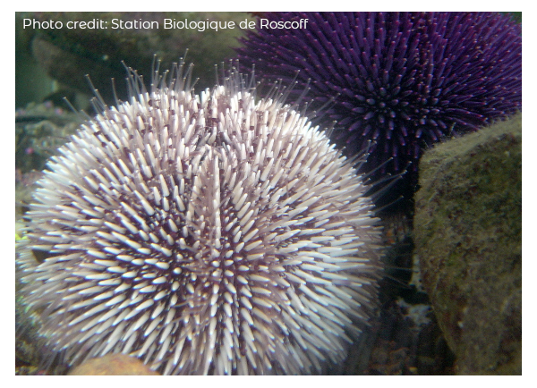
Welcome to the Echinoderms module. Sea urchins, particularly their gametes, have been an important experimental model since the end of the 19th century and throughout the 20th century. From Aristotle’s description of sea urchins’ feeding apparatus (350 BCE) to the genome sequencing of Strongylocentrotus purpuratus in the 21st century, echinoderms, on many occasions, have been used for deciphering fascinating processes in cell and developmental biology. You may find some description of the main discoveries using sea urchin in “Pontheaux F., Roch F., Morales J., Cormier P. (2021) Chapter 17 - Echinoderms: Focus on the sea urchin model In: Boutet, A. & B. Schierwater, eds. Handbook of Established and Emerging Marine Model Organisms in Experimental Biology, CRC Press.”
How to perform fertilizations of sea urchins
In this second video, you will learn how to handle adult sea urchins and induce the release of their gametes. You will also see how to carry out in vitro fertilization. This video is very close to what you are expected to do during the practical work.
Sea urchins, developmental biology, and GRNs


Next, in the two following interviews, you will find the personal views of two researchers on the use of this model in a more developmental and evolutionary context. Jenifer Croce and Smadar Ben-Tabou de Leon will tell you about their scientific journey and human encounters that have brought them further in their understanding of the mechanisms that control the regulation of genes during embryonic development in echinoderms.
The sea urchin and developmental biology, by Jenifer Croce (interview, PDF)
How a physicist becomes a GRN expert, by Smadar Ben-Tabou de Leon (interview, PDF)
How to monitor in vivo Cyclin B-CDK1 activity following fertilization
This third video focuses on how to monitor the activity of a key actor of the cell cycle. We show you how to set up an experiment, making it possible to measure in vivo the mitotic CDK1-cyclin B complex activity. We might think about how to adapt this protocol with other models or embryonic developmental stages during the practical.

“SUnSET” in sea urchins
Microinjection to measure mRNA translation efficiency
The fifth video is intended to show you how it is possible to measure the translational efficiency of a specific mRNA sequence. This kind of experiment requires producing molecular mRNA constructs allowing the translation of reporter gene under the control of an untranslated region and skillful approaches of microinjection into the eggs. The demonstration is performed by Florian, a 3rd-year Phd student (click here for Florian’s ResearchGate profile)
We are looking forward to meeting you in the in-person class, and we will be happy to answer whatever questions you may have.
For further reading, the following link leads to the Station Biologique de Roscoff where you can find our team’s recent publications: http://www.sb-roscoff.fr/en/team-translation-cell-cycle-and-development/publications
You can also find all the genomic data available on echinoderms on the "Echinobase" site at the following address: https://www.echinobase.org/entry/
